Caractéristiques :
- Solide, résilient, résiste à l’écaillage et à l’effritement.
- Libère et recharge du calcium, phosphate et fluor
- Liaison chimique - Élimine les micro-infiltrations
- Aucune sensibilité
- Hydrophile - Technique simplifiée
Description :
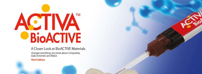
ACTIVA BioACTIVE - FOND DE CAVITÉ est le premier matériau de fond de cavité bioactif qui combine des propriétés d’absorption de chocs, de la force et de la durabilité. La liaison et l’intégration chimique à la dent supprimeront toute microfuite bactérienne avec une grande facilité d’application. ACTIVA BioACTIVE - Fond de cavité adhère chimiquement à la dentine, pour devenir partie intégrante de la dent. Il est également plus dur qu’un composite Flow et libère plus de fluor qu’un verre ionomère tout en fortifiant les dents. Après polymérisation, ACTIVA BioACTIVE - Fond de cavité devient dur et étanche contre l’infiltration bactérienne tout en réduisant considérablement les problèmes de sensibilité. Il est compatible avec toutes techniques de restauration.
Références :
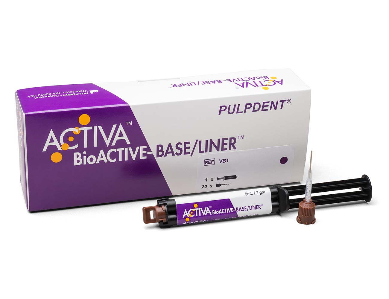
VB1 – Seringue de 5 ml / 7 g + 20 embouts (A20N1)
Teinte : Dentine
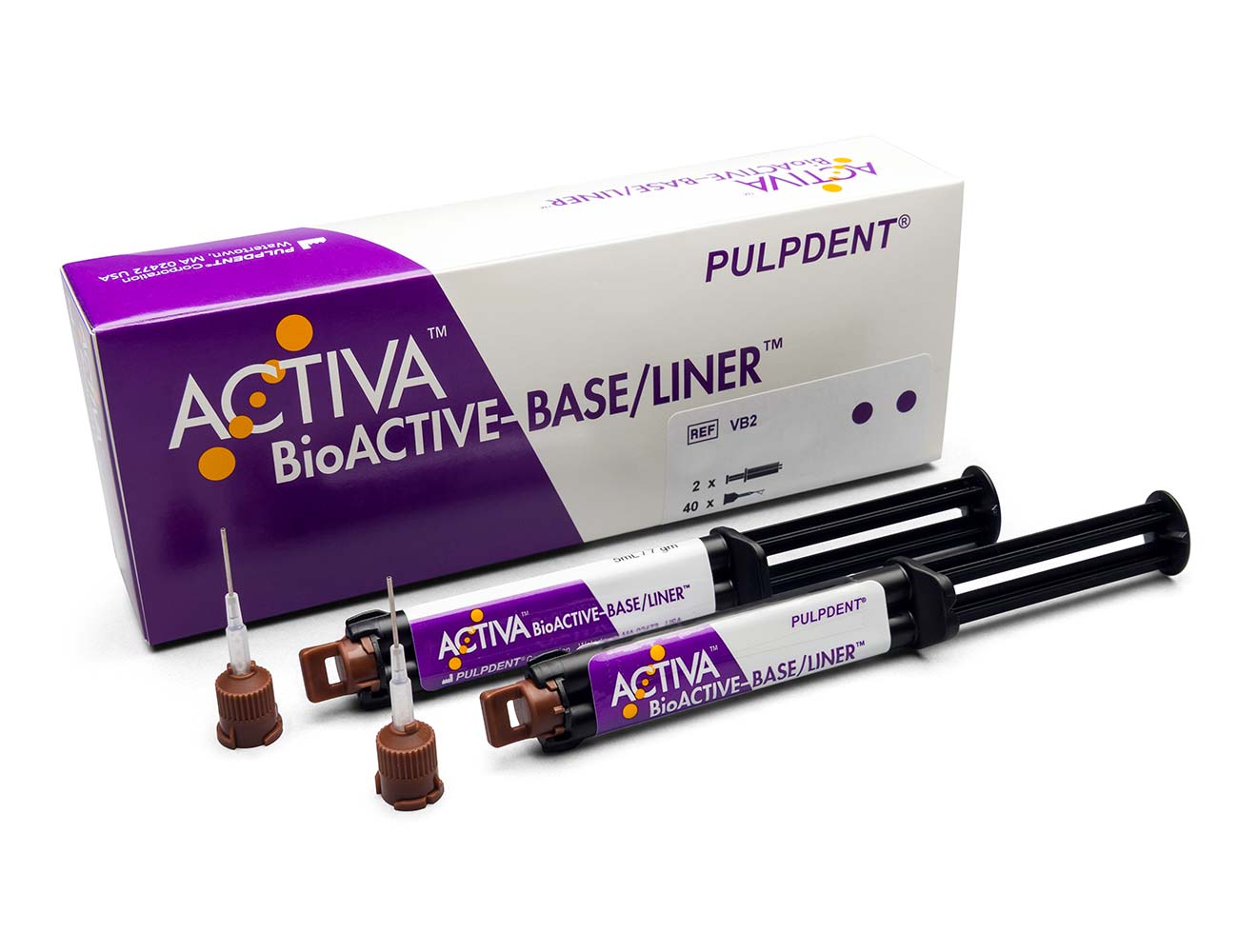
VB2 – 2 seringues de 5 ml / 7 g + 40 embouts (A20N1)
Teinte : Dentine
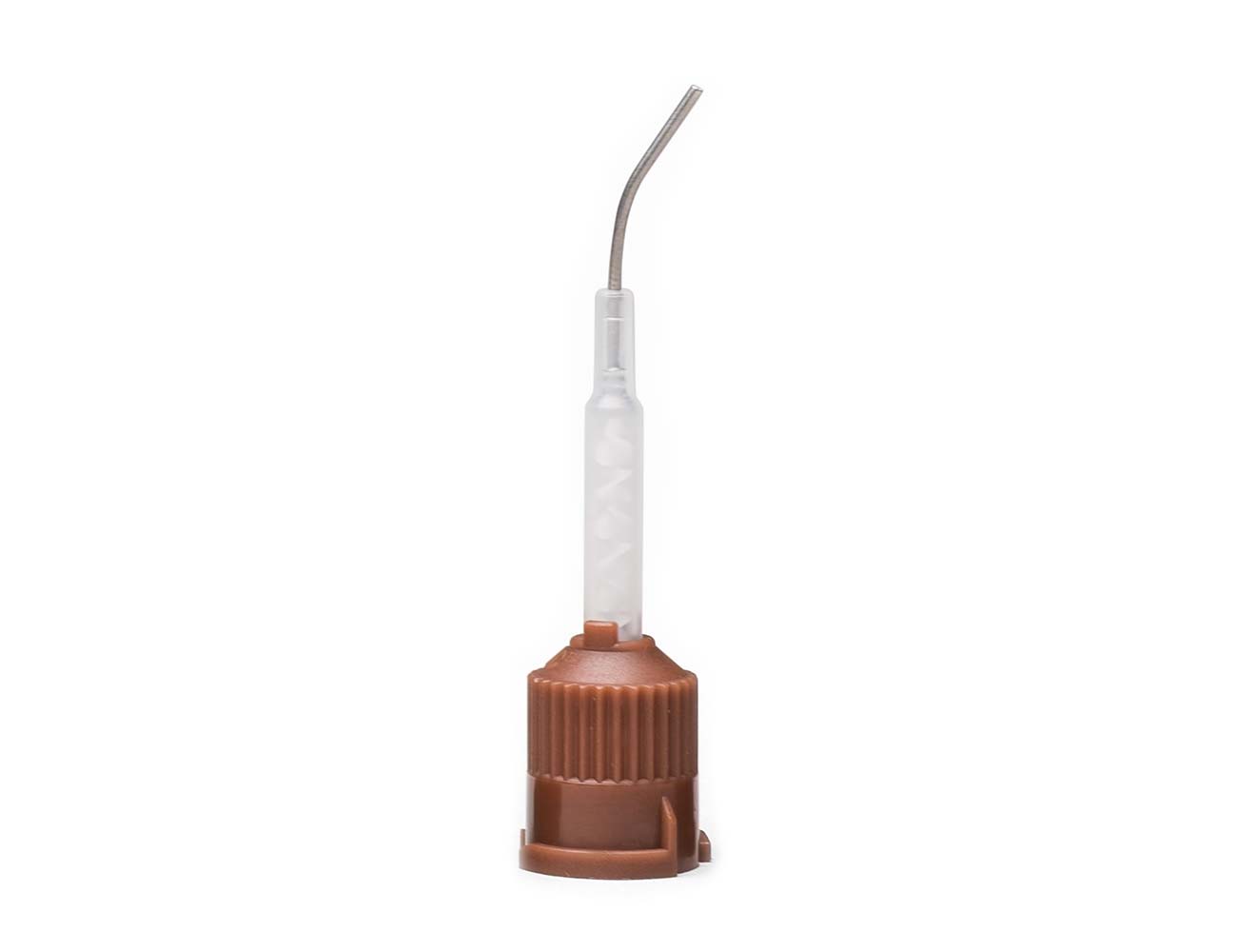
A20N1 – 20 embouts Automix directionnels, 20 ga

A50N1 – 50 embouts Automix directionnels, 20 ga
Propriétés Physiques
Temps de travail à température ambiante :
90 seconds
Temps de photopolymérisation :
20 secondes
Temps d’autopolymérisation à 37°C:
≤5 minutes
Pourcentage du filler (poids) :
45%
Pourcentage de verre ionomère (poids) :
15%
Libération de fluor cumulée à 7 jours :
23.8 µg/cm²
Résistance à la flexion :
86 MPa / 12 470 Psi
Résistance à la compression :
226 MPa / 32 770 Psi
Résistance à la pression diamétrale :
37 MPa / 5365 Psi
Épaisseur du film :
11 microns
Application
Activa Fond de Cavité est idéal pour les préparations profondes. Il fournit un réservoir d’ions de calcium, de phosphate et de fluor qui scelle et protège la dentine.
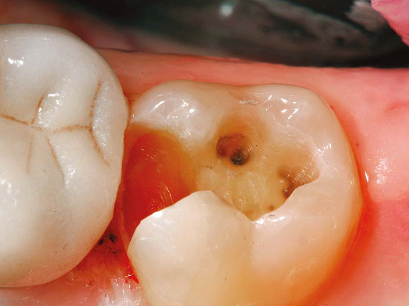
Fig. 1: Préparer la dent.
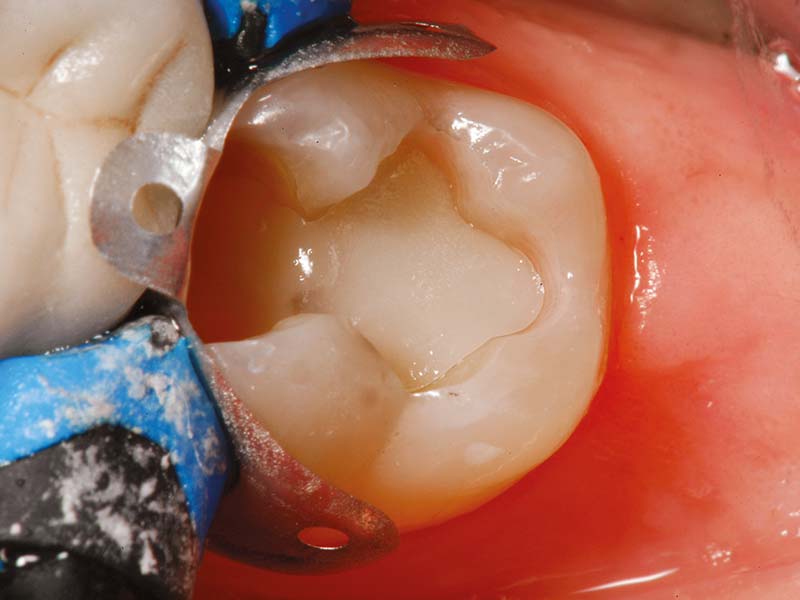
Fig. 2: ACTIVA BioACTIVE-Fond de cavité après polymérisation.
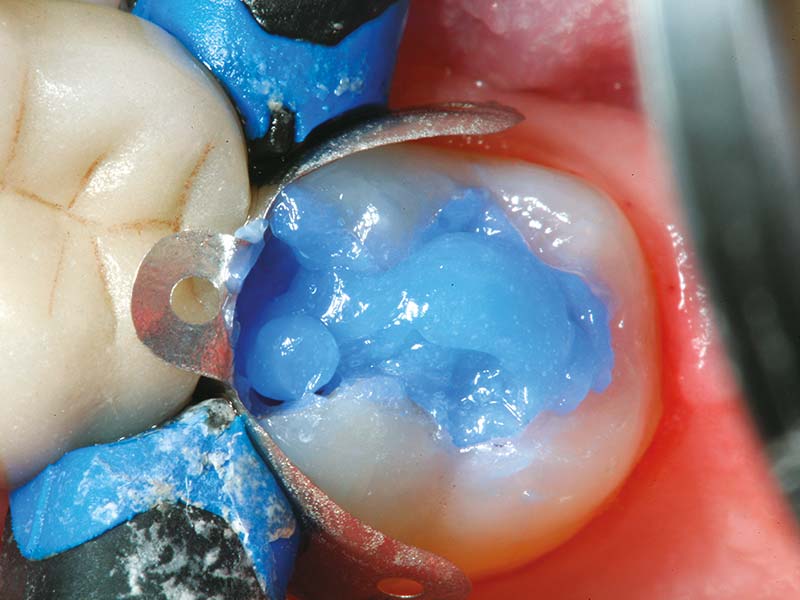
Fig. 3: Appliquer Etch-Rite pendant 5 secondes.
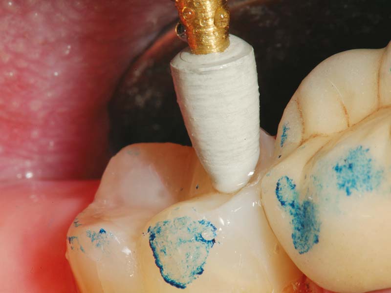
Fig. 4: Finir la restauration en utilisant ACTIVA BioACTIVE-Restauration.
Photos du Dr. Robert Lowe
Articles et Références
- Fluoride ion release and recharge over time in three restoratives. Slowokowski L, et al. J Dent Res 93 (Spec Iss A_ 268, 2014 (iadr.org).
- Zmener O, Pameijer CH, Hernandez S. Resistance against bacterial leakage of four luting agents used for cementation of complete cast crowns. Am J Dent 2014;27(1):51-55.
- Zmener O, Pameijer CHH, et al. Marginal bacterial leakage in class I cavities filled with a new resin-modified glass ionomer restorative material. 2013.
- Flexural strength and fatigue of new Activa RMGIs. Garcia-Godoy F, et al. J Dent Res 93 (Spec Iss A)_ 254, 2014 (iadr.org).
- Deflection at break of restorative materials. University testing. Submitted for publication.
- McCabe JF, et al. Smart Materials in Dentistry. Aust Dent J 201156 Suppl 1:3-10.
- Cannon M, et al. Pilot study to measure fluoride ion penetration of hydrophilic sealant. AADR Annual Meeting 2010.
- Water absorption properties of four resin-modified glass ionomer base/liner materials. (Pulpdent)
- pH dependence on the phosphate release of Activa ionic materials. (Pulpdent)
- Kane B, et al. Sealant adaptation and penetration into occlusal fissures. Am J Dent 2009;22(2):89-91.
- Rusin RP, et al. Ion release from a new protective coating. AADR Annual Meeting 2011.
- Sharma S, Kugel G, et al. Comparison of antimicrobial properties of sealants and amalgam. IADR Annual Meeting 2008. (iadr.org).
- Naorungroj S, et al.Antibacterial surface properties of fluoride-containing resin-based sealants. J Dent 2010.
- Prabhakar AR, et al. Comparative evaluation of the length of resin tags, viscosity and microleakage of pit and fissure sealants – an in vitro scanning electron microscope study. Contemp Clin Dent 2011;2(4):324-30.
- Pameijer CH. Microleakage of four experimental resin modified glass ionomer restorative materials. April 2011.
- Microleakage of dental bulk fill, conventional and self-adhesive composites. Cannavo M, et al. J Dent Res 93 (Spec Iss A) 847, 2014 (iadr.org).
- Comparison of Mechanical Properties of Dental Restorative Material. Girn V, et al. J Dent Res 93 (Spec Iss A) 1163, 2014 (iadr.org).
- Mechanical properties of four photo-polymerizable resin-modified base/liner materials. (Pulpdent)
- Singla R, et al. Comparative evaluation of traditional and self-priming hydrophilic resin. J Conserv Dent 2012;15(3):233-6.
- Water absorption and solubility of restorative materials. (Pulpdent)
- www.nidrc.nih.gov
- Spencer P, et al. Adhesive/dentin interface: the weak link in the composite restoration. Am Biomed Eng 2010;38(6):1989-2003.
- Murray PE,et al. Analysis of pulpal reactions to restorative procedures, materials, pulp capping, and future therapies. Crit Rev Oral Biol Med 2002;13:509.
- DeRouen TA, et al. Neurobehavioral effects of dental amalgam in children: a randomized clinical trial. JAMA 2006;295(15):1784-1792.
- Nordbo H, et al. Saucer-shaped cavity preparations for posterior approximal resin composite restorations: observations up to 10 years. Quintessence Int 1998;29(1):5-11.
- Skartveit L, et al. In vivo fluoride uptake in enamel and dentin from fluoride-containing materials. J Dent Child 1990; 57(2):97-100.
- Wear of calcium, phosphate and fluoride releasing restorative material. University testing. Submitted for publication: IADR 2015. (iadr.org).
- Surface roughness and wear resistance of ACTIVA compared to glass ionomers, RMGIs and flowable composites. University testing. Submitted for publication: IADR 2015. (iadr.org).


